Figure 2
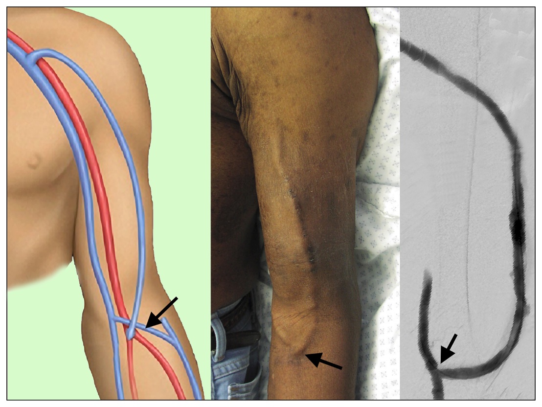
Images of a brachial-cephalic AVF drawing of surgical anatomy (left panel), a matured ready and used for dialysis cannulation (middle panel), and the contrast angiographic appearance (right panel)
Images of a brachial-cephalic AVF drawing of surgical anatomy (left panel), a matured ready and used for dialysis cannulation (middle panel), and the contrast angiographic appearance (right panel)